Analysis of building blocks of biomolecules: determination of the protonation site
![]() |
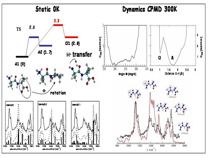
IRMPD offers a direct possibility for determination of protonation sites by comparison between experimental IR absorption spectrum and the predicted spectra of the different tautomers. In the case of protonated dialanine, static calculations of the lowest energy configurations show that the proton can be located at the N-terninal (labelled A1 and A2) or C=O (labelled O1) amide groups at 300 K. These conformations are separated by energy barriers with transition states accessible at room temperature. Conformations A1 and A2 give the best agreement with the experimental spectrum while conformation O1 contributes to a much less extent to the experimental spectrum. Car-Parrinello molecular dynamics (CPMD) simulations on Ala-AlaH+ peptide at 300K have shown a continual and recurrent isomerization dynamics between the two A1 and A2 conformers, i.e. the internal energy of the peptide at 300K is sufficient to overcome the energy barrier separating the A1 and A2 basins on the potential energy surface. Besides, one important result is the observation of spontaneous and reversible proton transfer events between NH3+ (A conformer) and the adjacent C=O amide (O conformer). This indicates that O1 is a metastable state which is thermally accessible at 300K. The IR spectrum extracted from the CPMD simulation is calculated from the Fourier Transform of the dipole time correlation function. All pertinent band shaping effects, such as anharmonicity and temperature are incorporated ab inito via the CPMD methodology. |
Gas phase structure of flexible peptide : Arginine-Glycine-Aspartic acid (RGD)
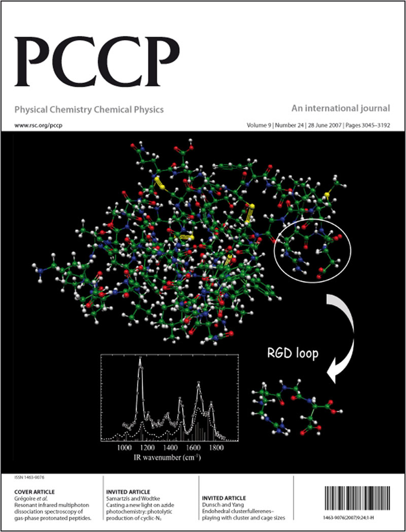
An important recognized motif involved in cell adhesion and inflammatory processes is the arginine-glycine-aspartic acid RGD sequence, found in large glycoproteins such as fibronectin, participating to the composition of the intercellular medium. It is specially recognized by transmembrane proteins called integrins and initiates cell-signalling processes. We have shown that the protonated RGD sequence adopts in the gas phase at least two conformations at 300K, one being similar to the structure of RGD as embedded in a protein (dendroaspin). In this neurotoxin homologue, the flexible RGD loop structure corresponds to residues 43-45 located on the surface of the protein. Our gas-phase study has shown that this loop structure is an intrinsic structural property of RGD, even if the acid aspartic carboxyl group is neutral while it is deprotonated for peptides in solution. |
![]() |
Transition between gas-phase and aqueous solution structures of a neurotransmitter
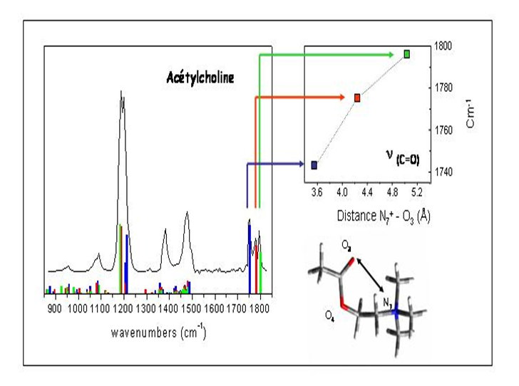
Molecular recognition of acetylcholine and its agonists by nicotinic or muscarinic acetylcholine receptors is a widely studied systems due to its importance in memory, cognition and reward processes as well as its potential implications in the treatment in Alzheimer's and Parkinson's diseases. Among the key elements for biorecognition of these molecules in the binding sites of the receptors, called "pharmacophores", are a cation-&pi interaction and an accepting hydrogen bond interaction with receptor residues. The distance between these two pharmacophoric sites is thus a crucial parameter that is usually invoked for explaining the strong biochemical similarities observed between acetylcholine and either nicotine and muscarine ions.
![]() |
Structural information on acetylcholine and its two agonists, nicotine and muscarine has been obtained from the interpretation of IR spectra recorded in the gas-phase (IRMPD) or in low pH aqueous solutions (FT-IR). In the gas-phase, the IRMPD spectrum of acetylcholine ions exhibits three distinct transitions of the carbonyl stretch around 1750 cm-1, which implies that at least three different conformations are populated at 300K. Among these conformers, only one corresponds to a N+-O distance (5.05 Å) that fits within the bioactive range, while the four others low energy conformers exhibit much shorter N+-O distance. However, in aqueous solution, this distance in the four lowest energy conformations is comprised in between 4.6 and 5.2 Å, all compatible with the bioactive range. The conformational space of these very flexible molecules is thus strongly modified by the presence of the solvent. |
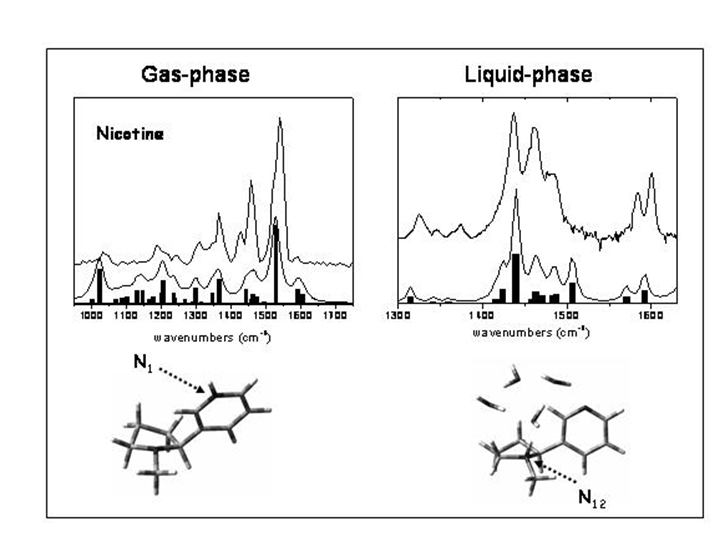
In the case of nicotine, the solvatation induces a complete change of the structure and the protonation site. Nicotine possesses two protonation sites on the nitrogen of the pyridine (labelled N1(sp2)) and pyrrolidine (labelled N12(sp3)) cycles. In solution, a strong transition at 1450 cm-1 is observed and corresponds to the bending mode of N12-H+. In the gas phase, the IRMPD spectrum of protonated nicotine exhibits an intense transition centred at 1540 cm-1 that involves the bending motion of N1-H+ group. This result has provided for the first time a clear experimental evidence for protonation of nicotine on the pyridine site, which is not the bioactive site of nicotine in condensed phase. |
![]() |
Structural characterization of large biomolecules: DNA strands containing G-quadruplex
![]() |
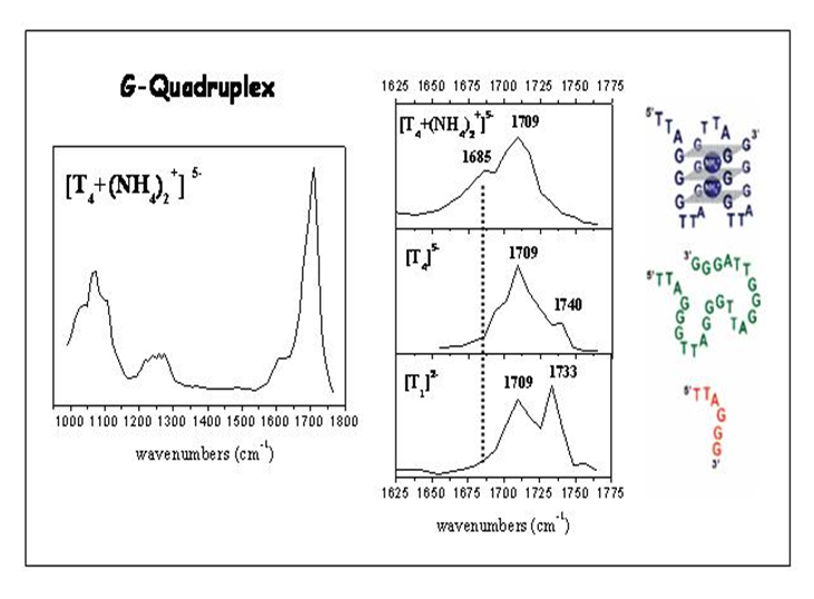
Nucleic acid secondary structures are determined by hydrogen bonding interactions between nucleic bases. Besides the well-known Watson-Crick base pairing motif, there is a variety of other possible hydrogen bonding configurations. For instance, guanine-rich sequences can fold into G-quadruplex structures owing to Hoogsteen hydrogen bonding between four guanines, forming G-quartets. As evidence is accumulating on in vivo G-quadruplex formation in telomeric DNA and in the promoters of some oncogenes, these structures have become key targets for anticancer strategies. G-quadruplexes are present in telomeres (ends of human chromosomes) that consist of long DNA single strands containing repeats of TTAGGG sequence. We have investigated the IR signature of two quadruplex-forming sequences: dTG4T, forming a tetrameric quadruplex [(dTG4T)4(NH4+)3] that is highly stable in solution and in the gas phase, and the human telomeric sequence dTTAGGGTTAGGGTTAGGGTTAGGG (noted T4), forming an intramolecular antiparallel quadruplex in ammonium acetate solution. For both species, a significant red shift of the C=O stretch mode of the guanine base is observed for the non-covalent complexes with ammonium cations sitting in between two successive G-quartets. This first experimental study of large biomolecules has proven the ability of IRMPD spectroscopy for gas-phase conformational analysis of rather complex systems. |
Molecular recognition in biomolecular non-covalent complexes
Vancomycin is a naturally occurring glycopeptide antibiotic active against Gram-positive bacteria and is considered as a drug of last resort. Its original use is for the treatment of penicillin-resistant Staphylococcus aureus. Vancomycin binds to the bacteria cell-wall peptidoglycan precursor through noncovalent interactions that inhibit the action of specific bacterial enzymes involved in the development of the cell wall through cross-linking with pentaglycine, leading to its cleavage and finally the death of the bacteria. Early studies have shown that vancomycin binds efficiently to the peptidoglycan precursor UDP-N-acetylmuramyl-LA-DE-LK-DA-DA with a high affinity of 1.6x10⁵ M-1, while further works have demonstrated that the essential region of this peptide responsible for the formation of the complex is the DAlanyl-DAlanine terminus sequence.
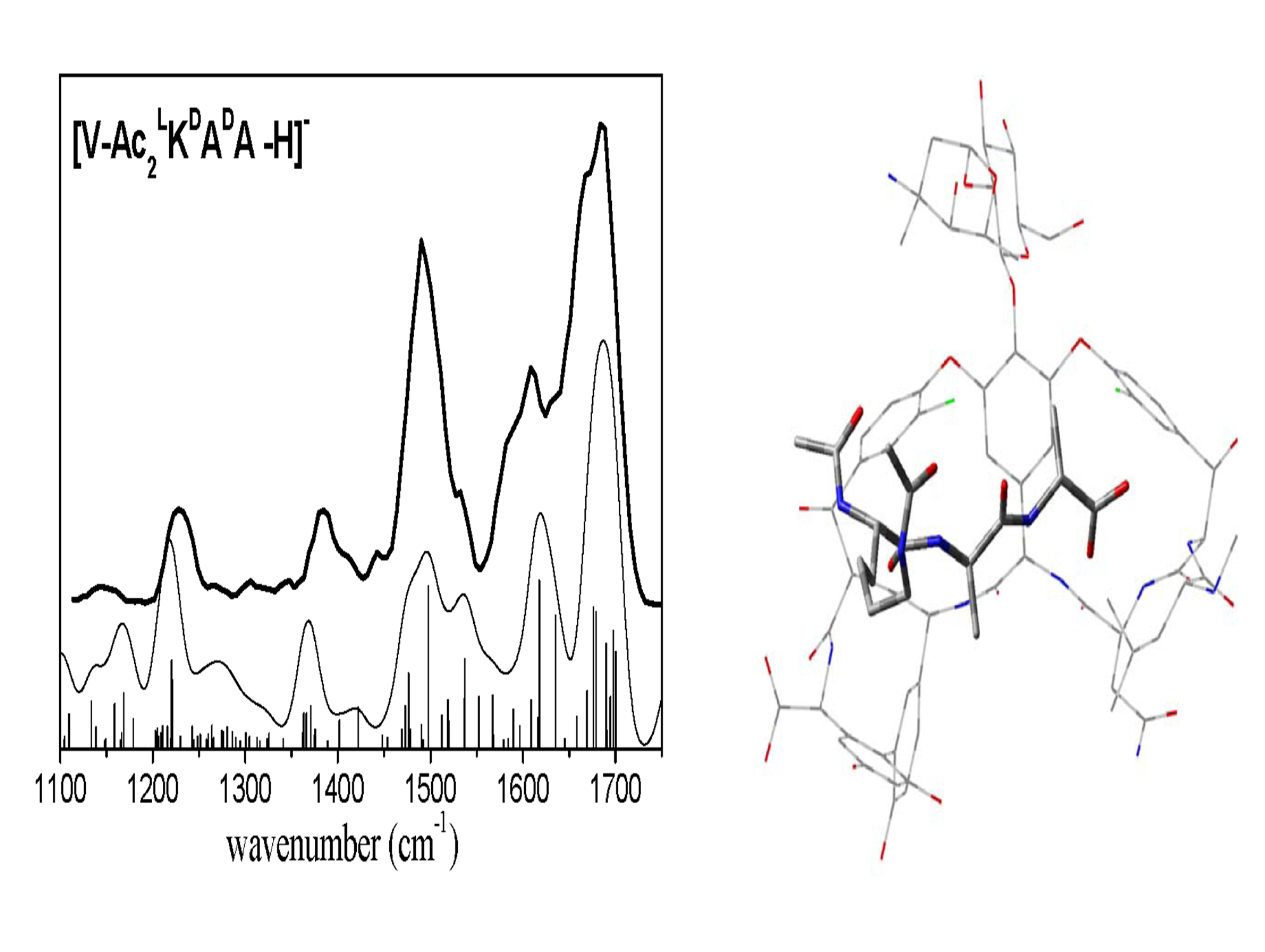
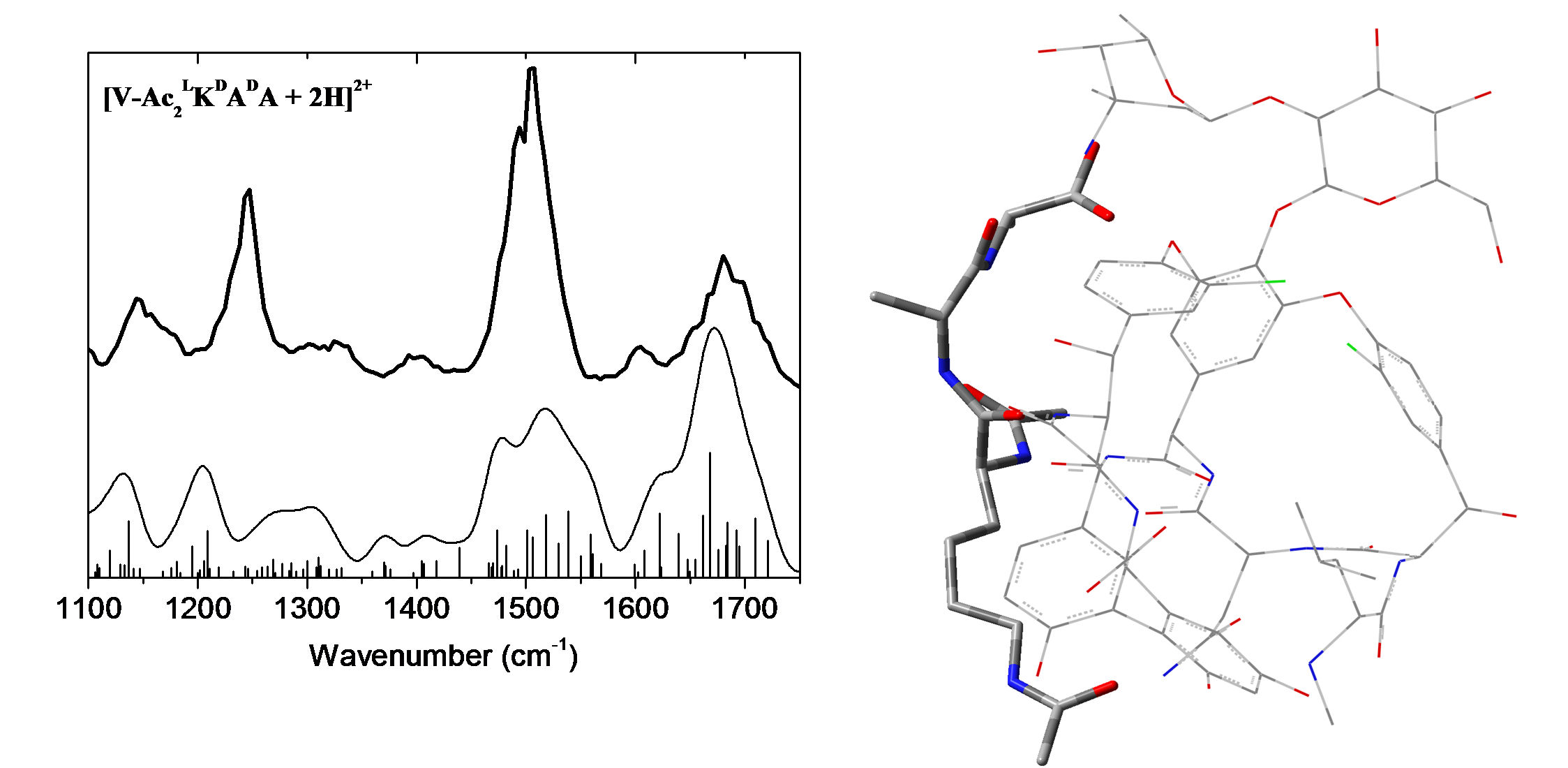
The binding of vancomycin to the tripeptide cell-wall precursor analogue Ac2LKDADA involves several intermolecular hydrogen bonds, three with the carboxylate group of the tripeptide and two between amide groups of both species. The binding pocket thus contains the pseudopeptidic chain of vancomycin while the benzoic cycles and the sugar moiety do not seem to be directly involved in the formation of the complex. The mid-IR spectra (1100-1800 cm-1) of these mass-selected anionic species have been recorded by means of resonant infrared multiphoton dissociation (IRMPD) spectroscopy performed with the free-electron laser CLIO. Structural assignment has been achieved through comparisons with the low-energy conformers obtained from replica-exchange molecular dynamics simulations, for which IR spectra were calculated using a hybrid quantum mechanics/semi-empirical (QM/SE) method at the DFT/B3LYP/6-31+G*/AM1 level. Comparison between deprotonated vancomycin and its non-covalently bound V+Ac2LKDADA complex shows significant spectral shifts of the carboxylate stretches and the Amide I and Amide II modes that are satisfactorily reproduced by the structures known from the condensed phase. |
![]() |
![]() |
The situation is rather different in the protonated form. Neutralization of the carboxylate group impairs the specific interaction of the deprotonated group of the receptor with vancomycin. Earlier gas-phase studies have suggested that the native structure of the complex might not be preserved in the protonated form, although no direct spectroscopic study has been yet undertaken. The structure of doubly protonated vancomycin antibiotics with its cell-wall precursor analogue Ac2LKDADA has been investigated in the gas phase through a combined laser spectroscopy, ion mobility (in collaboration with the group of Ph. Dugourd, LASIM) and theoretical modeling approach. Replica-exchange molecular dynamics simulations using the Amber99 force field were performed to explore the potential energy landscape of isolated vancomycin ions, as well as the different binding sites with the receptor. Among the low-energy conformers found, those with a calculated diffusion cross-section consistent with ion mobility experiments were selected for further optimization, and their IR spectra were simulated using a hybrid quantum mechanics/semi empirical (QM/SE) method at the DFT/B3LYP/6-31g(d):AM1 level. Both theoretical and experimental findings provide strong evidence that the native structure of the complex is not preserved in vacuo for the doubly protonated species. |